New customers save 5% with the code GET05
-
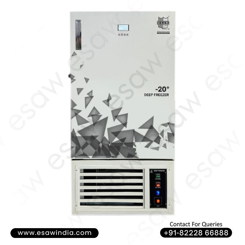
-20°C Laboratory Deep Freezer
Regular price Rs. 195,990.00 -
-25°C Laboratory Deep Freezer
Regular price Rs. 195,000.00 -
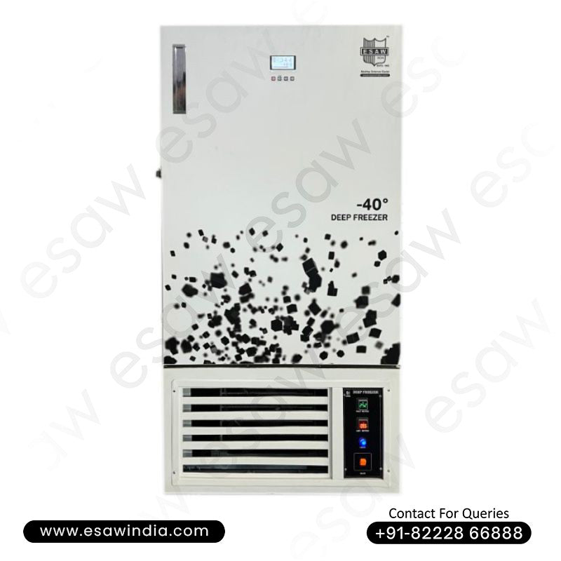
-40°C Laboratory Deep Freezer
Regular price Rs. 185,000.00 -
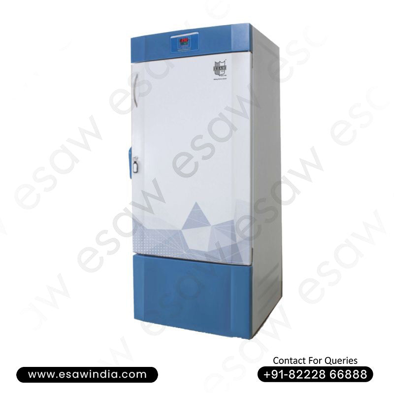
-80°C Laboratory Deep Freezer
Regular price Rs. 197,000.00